Princeton Radiology Collaborates with Local Urologists to Offer The Next Generation in Prostate Care
Princeton Radiology has acquired UroNav software by Invivo. With this new software we are able to gather pre-biopsy MRI images of the prostate and then seamlessly transfer them to local Urologists. Using UroNav, these Urologists can fuse the pre-biopsy MRI images with real time ultrasound-guided biopsy images for excellent delineation of the prostate and suspicious lesions, as well as clear visualization of the biopsy needle – allowing Urologists to directly aim biopsy needed at lesions. Better images means better results.
Prostate cancer is the only type of solid organ tumor that is usually diagnosed sight unseen with hit-or-miss tissue biopsies.
For decades, Urologists have used a systematic, but blind approach to sample prostate tissue in men with an elevated PSA level and no palpable lesion, taking up to 18 core needle biopsies in scattered sections of the organ. Using this method doctors can’t be sure that they haven’t missed an aggressive tumor hidden in the 99% of tissue that was not biopsied, and they sometimes end up sticking needles into men who have no tumors, only an elevated PSA level.
Now, due to the precise targeting provided by MRI, many fewer biopsy samples are needed for accurate diagnosis – sometimes 80 to 90% fewer. Fewer needle-sticks means lower risk of infection, bleeding, pain, and means and shorter recovery time. And thanks to the recent advance called multi-parametric MRI, specially trained radiologists can gauge the aggressiveness of a prostate lesion not only by how it looks, but also by how tightly its cells are packed, how blood flows through it, and its chemical makeup.
Targeted MRI/Ultrasound biopsy is poised to become the new standard in prostate cancer detection. Princeton Radiology is proud to collaborate with local Urologists to bring the next generation of prostate care to our patients.
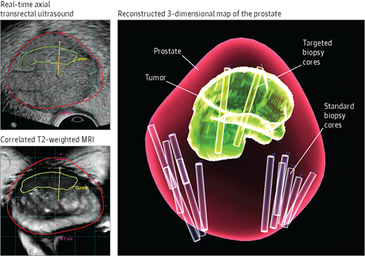
Image: Magnetic resonance (MR)/ultrasound fusion combines MR images of the prostate (bottom left, red line) with real-time ultrasound images of the prostate (top left, red line) to assist in targeted biopsy of a previously identified lesion (green line). The location of the biopsy can be recorded (yellow line), and a reconstructed 3-dimensional map of the prostate can be generated at the conclusion of the biopsy (right). Standard biopsy cores and targeted biopsy cores are highlighted here for comparison.