Women's Services
Princeton Radiology is dedicated to empowering women with comprehensive screenings and information essential for safeguarding their health through every life stage, from child-bearing to menopause. Our array of screening, diagnostic, and monitoring tests ensures a proactive approach to women’s health.
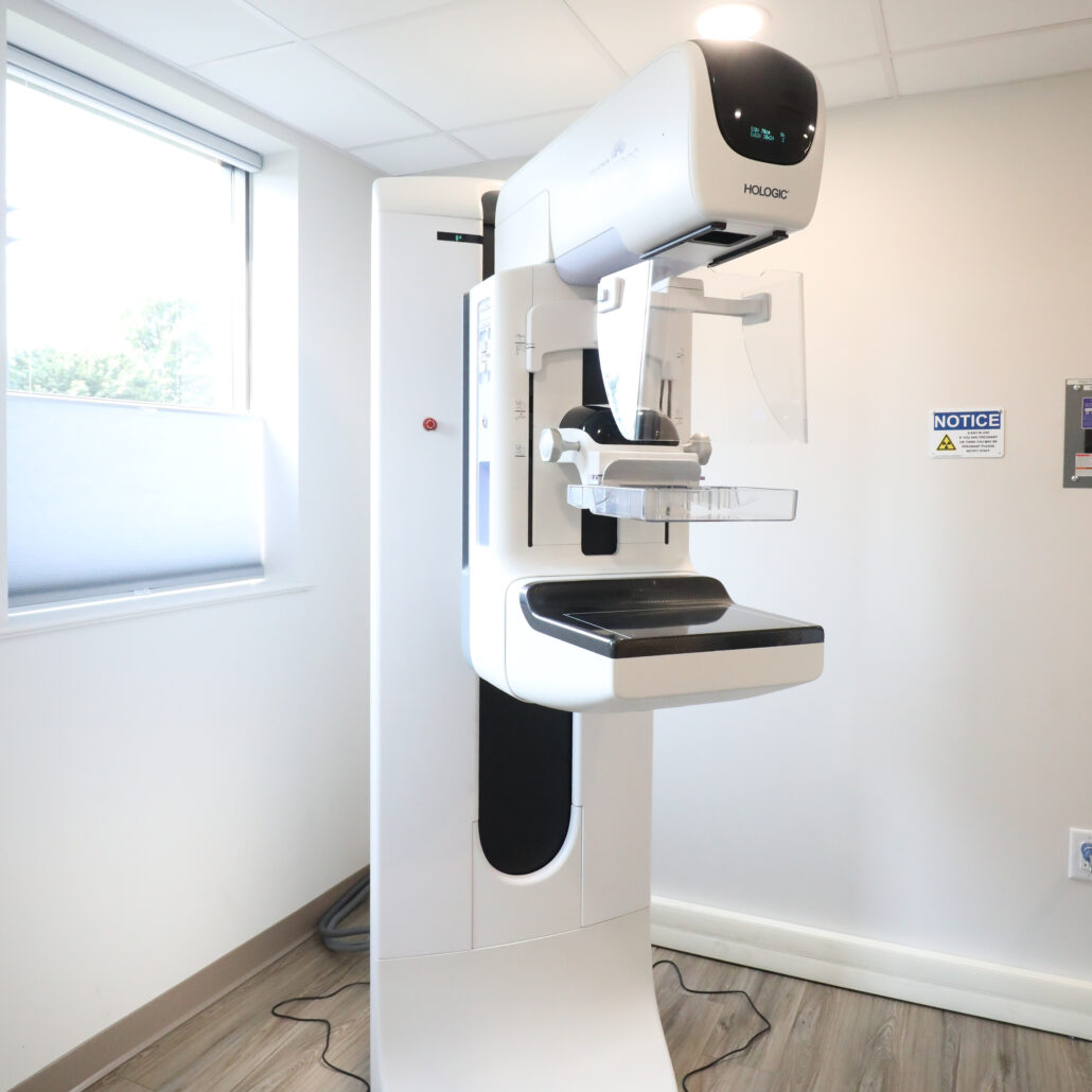
Digital Mammography in New Jersey and Newtown, PA—Now with Heart Health Insights
Digital Mammography stands out as the most effective method for early breast cancer detection, and at Princeton Radiology, we elevate the standard with 3D SmartMamm®—a proactive approach to breast health. Our team assesses your lifetime risk of breast cancer by calculating your risk score, so you and your doctor can create a customized plan to properly monitor your breast health. Yearly screening mammograms are recommended for women over the age of 40. During a digital mammogram, breasts are gently compressed so detailed computerized images can be taken to help identify any unusual tissue. SmartMamm now also offers heart health insights with reporting on breast arterial calcifications at no additional charge.

Breast MRI in New Jersey and Newtown, PA
SmartBreast™ MRI serves as a supplemental tool for an in-depth examination of suspicious breast tissue. Using a magnetic field, the MRI scanner produces detailed images of the breasts’ interior, aiding in accurate diagnosis.
Bone Density (DEXA) in New Jersey and Newtown, PA
The Bone Density (DEXA) helps women monitor bone loss and detect the early symptoms of osteoporosis. Osteoporosis can result in chronic pain and bone fractures. With early detection, osteoporosis can be treated before debilitating fractures occur. The DEXA (Dual Energy X-Ray Absorptiometry) scan is quick and painless.


Body Composition Analysis (DXA) in New Jersey and Newtown, PA
DXA is a valuable tool for physicians, athletic trainers and fitness enthusiasts. It precisely measures the amount of fat, muscle and bone in your body. This information helps physicians and clinicians measure and manage obesity, evaluate candidates for bariatric surgery, monitor retention of lean muscle mass for patients taking GLP-1 medications, and evaluate weight management programs. It is also helpful in assessing disease risk, as well as muscle loss and growth disorders.