DEXA Scan Bone Densitometry
At Princeton Radiology, we make it easy to take a proactive step in protecting your bone health. Our advanced Bone Densitometry (DEXA) scans offer fast, accurate assessments to detect osteoporosis and track bone loss—often before symptoms appear. With expert radiologists and leading-edge technology, we provide the clarity you need to stay strong and supported at every stage of life.
What is a DEXA scan?
Bone Densitometry is a fast, safe and painless test that uses advanced technology called DEXA (Dual Energy X-Ray Absorptiometry) to measure symptoms of osteoporosis — such as low density and mineral content of bone — that may have developed unnoticed over many years. Because osteoporosis can result in bone fractures that can cause chronic pain, disability and loss of independence, it is important to begin treating osteoporosis at an early stage. Bone densitometry can detect the early signs of osteoporosis so that patients can begin treating it before a debilitating fracture occurs.
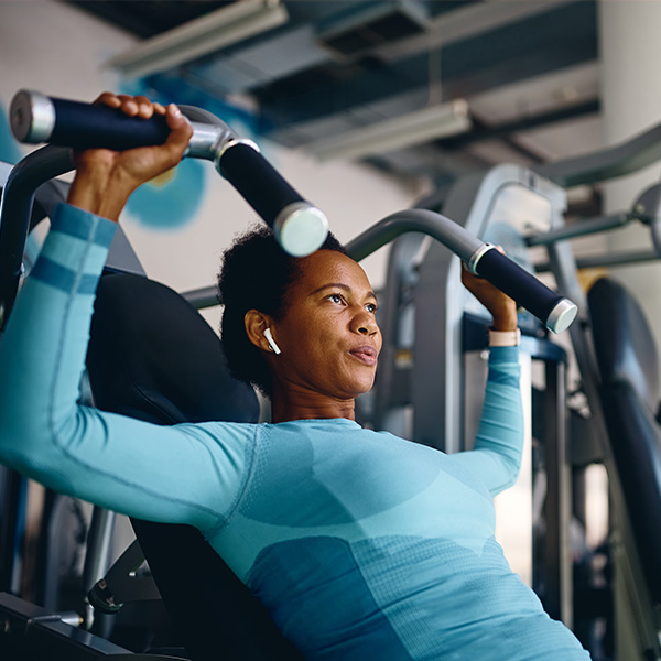
Physicians and clinicians use DEXA Body Composition Analysis to help assess and manage obesity. It’s also helpful in assessing age-related muscle loss, neuromuscular changes, and growth disorders. Knowing your FMI can help your clinician plan and monitor interventions for excessive body fat, and track the progress of physical training regimens. DEXA Body Composition Analysis may also benefit people who are:
- Diabetic
- Candidates for bariatric surgery or receiving follow-up care after a procedure
- Suffering from sarcopenia (degenerative muscle loss)
- Currently involved in a weight management program
- Anorexic
- Fitness enthusiasts
Please be sure to bring your referral from your doctor with you when you come for your appointment. If you do not have it with you, we may not be able to perform your test.
Please arrive 30 minutes early to register.
During a comprehensive DXA bone evaluation, a patient lies comfortably on a padded table while the DEXA unit scans one or more areas of his/her body, usually the spine or hip because they are particularly prone to fracturing.
When the Bone Densitometry exam is complete, your images are sent to a computer and analyzed. They are then given to a radiologist, a physician who specializes in the diagnostic interpretation of medical images. After your Bone Densitometry study has been reviewed, your personal physician will receive a report of the findings. This report will include your bone mineral density (BMD), along with your FRAX results. The radiologist will use the FRAX assessment tool, developed by the World Health Organization, to obtain two results, expressed as percentages. These numbers are a ten-year probability of hip fracture and ten-year probability of a major osteoporotic fracture (clinical spine, forearm, hip or shoulder fracture).
This exam is available at the following locations:
Over 60 years of excellence in imaging.
And that’s just one reason why Princeton Radiology is the right choice.
Interpretations by board certified, sub-specialty trained radiologists.
Fast scheduling, less time in the waiting room, and same-day or next-day results in most cases.
We cater to patient schedules with daytime, evening and weekend hours for many exams.
From our friendly and caring staff to our beautiful imaging facilities, everything we do is patient-focused.