IDEAS Study (Alzheimer’s)

Princeton Radiology Participated in Largest Brain Amyloid Scan Research Study

Princeton Radiology is a registered PET imaging site for the Imaging Dementia – Evidence for Amyloid Scanning (IDEAS) study

sponsored by the Alzheimer’s Association and managed by the American College of Radiology (ACR) and American College of Radiology Imaging Network (ACRIN) from February 2016 to the study’s conclusion in December 2017. Princeton Radiology offered Medicare-covered amyloid PET scans to study eligible Medicare patients who were referred by participating IDEAS dementia specialists (neurologists, psychiatrists and geriatric medicine physicians). The research study sought to determine the clinical usefulness of PET imaging of brain amyloid deposits in the diagnosis of patients who are being evaluated for Alzheimer’s disease and dementia.

Diagnosing Alzheimer’s disease is complex. With no single test currently available, diagnosis is based on an individual’s history, physical examination and cognitive testing. Amyloid PET imaging represents a potential major advance in the assessment of people with cognitive impairment. The scan visualizes plaques present in the brain that are prime suspects in damaging and killing nerve cells in Alzheimer’s. Before amyloid PET, these plaques could only be detected by examining the brain at autopsy. The IDEAS study gathered data from Medicare beneficiaries to determine if amyloid PET scans help physicians to make more informed treatment decisions.

For more information about the IDEAS study, visit:  http://www.ideas-study.org/

Freehold Radiology is now a Princeton Radiology Practice

After thoughtful discussion between both practices on how our teams could best collaborate, Princeton Radiology merged with Freehold Radiology on October 23, 2017. The merger extends Princeton Radiology’s services into Monmouth County allowing us to provide more of Central New Jersey with exceptional diagnostic imaging and interventional radiology services.

We fully expect the merger to enhance our already-exemplary standards to still-higher levels by creating an even greater depth of subspecialty expertise, complementing our team with the skillset of Freehold Radiology’s distinguished physicians.

As a practice that has always been committed to striving for the best for our community, we look forward to the benefits this integration will provide to our patients and their referring physicians. In a word, our community can expect MORE—more expertise, more accessibility, more services, with:

  • Five office locations
  • 36 board certified radiologists
  • 90+ years of combined commitment to excellence
  • More appointment times, including evenings and weekends
  • More service offerings
  • More of the latest, most advanced technology
  • More participation with all major insurance plans

Over the coming months, we will be working to combine the two practices clinically, operationally, and culturally. You may notice some changes that reflect our expanded practice, but the excellent standard of service you have always received from Princeton Radiology will remain the same—enhanced by the expertise of more physicians.

In this energizing time of change, questions from patients are to be expected. Please contact our scheduling team at 609.921.8211 with any questions about the merger.

Thank you for the opportunity to have served your medical imaging needs in the past. We look forward to a bright future, collaborating with our new colleagues from Freehold Radiology to help build a healthier community by expanding access to Excellence in Imaging.

HEALTH MATTERS: Innovative Treatments Target Liver Cancer

Did you know that the incidence of primary liver cancer has more than tripled since 1980?

More than 40,000 Americans will be diagnosed with primary liver cancer this year, according to the American Cancer Society. And thousands of others will be diagnosed with secondary (metastatic) liver cancer, cancer that spreads to the liver after starting somewhere else in the body.

While surgery remains the gold standard for treating liver cancer, advances in medicine have made more treatment options available, especially for patients who may not be able to tolerate surgery or in cases where tumors are too large to be removed safely.

At the Edward & Marie Matthews Cancer Center at University Medical Center of Princeton, patients have access to a full range of services for the diagnosis and treatment of cancer, including liver cancer.

Liver Basics

Your liver is your largest internal organ and is positioned under your right ribs just beneath your right lung. It consists of two lobes, and weighs a total of about 3 pounds. It is also unique in that it has two blood supplies — the portal vein and the hepatic artery.

Your liver performs many vital functions including removing toxins from your blood, breaking down and storing nutrients that power your body and repair tissues, and producing bile to help your body absorb nutrients.

Many primary liver cancers are related to the hepatitis B or hepatitis C virus, which increase the risk for liver cancer. Other common risk factors include:

Gender. More men develop liver cancer than women.

Cirrhosis. A disease that causes scarring of the liver and is most associated with alcohol abuse and hepatitis, cirrhosis increases the risk for liver cancer.

Diabetes. Type 2 diabetes has been linked with an increased risk of liver cancer, usually in patients who have other risk factors such as heavy alcohol use, chronic viral hepatitis, or a combination of the two.

Obesity. Obesity can increase the risk for liver cancer as it is associated with diabetes as well as non-alcoholic fatty liver disease, which can lead to cirrhosis.

In addition, liver cancer can also be caused by other cancers that have started elsewhere in the body and metastasized to the liver. Metastatic liver cancer is most commonly associated with colon, lung and breast cancer.

Advances in Treatment

Traditional treatment for liver cancer typically involves surgery, chemotherapy and external beam radiation.

However, not all patients are candidates for surgery and some may not respond to traditional chemotherapy and radiation.

Today, there are several innovative approaches — such as radioembolization, chemoebolization and microwave ablation — that are making treatment more precise and more accessible to a broader number of patients.

With radioembolization, radiologists insert a catheter into the hepatic artery — the blood supply to the tumor — and insert tiny beads that lodge in the artery and give off small amounts of radiation, selectively targeting the tumor.

The procedure, which is performed through a small incision in the groin or the wrist and guided by X-ray images, kills the cancer cells and shrinks the tumor while sparing the surrounding healthy liver tissue.

Radioembolization is a generally a treatment option for cancer that is largely confined to the liver and for patients who meet certain baseline standards for liver function.

Similar to radioembolization, chemoembolization uses a catheter in the hepatic artery to delivery tiny beads of chemotherapy to the tumor. It is most beneficial in patients whose cancer is predominately limited to the liver.

Both radioembolization and chemoembolization block or reduce blood flow to the tumor, helping to kill off the cancer cells. Because the main blood supply to the liver is through the portal vein, healthy liver cells are relatively unharmed.

Another procedure, microwave ablation uses heat to destroy tumors. With microwave ablation, doctors — guided by an ultrasound or CT scan — insert a needle-like probe into the tumor. The probe emits heat from microwave energy that reaches temperatures greater than 100 degrees and kills the cancer cells.

Microwave ablation is typically used to treat smaller tumors, generally no more than 2 inches across.

All three procedures are generally well tolerated by patients. Side effects are typically mild and may include low-grade fever, abdominal pain and nausea. Usually, the procedures are performed on an outpatient basis.

In addition, because the procedures are precisely targeted there is normally little damage to healthy liver cells.

An Individualized Approach

As with most diseases, there is no single right approach for treating liver cancer. Each patient is different and requires a treatment plan that is specific and targeted to their unique condition.

Fortunately, innovations in cancer care are enabling doctors to manage complex cancer cases with more precise treatment, ultimately providing better outcomes for their patients.

If you have been diagnosed with liver cancer, talk with your doctor about your treatment options and the safest and most effective approach for your individual condition.

The Edward and Marie Mathews Cancer Center at UMCP is accredited with commendation from the American College of Surgeons’ Commission on Cancer and a recipient of the American College of Surgeons’ Commission on Cancer Outstanding Achievement Award. UMCP and the UMCP Breast Health Center earned accreditation from the National Accreditation Program for Breast Centers, and the UMCP Breast Health Center is designated as a Breast Imaging Center of Excellence by the American College of Radiology.

This article was written by our own Samuel J. Greene, M.D. and originally published on CentralJersey.com.

Updated DXA Policy for Medicare Patients

March 6, 2017

Medicare covers DXA Bone Densitometry for certain Medicare beneficiaries who fall into at least one of the following categories:

1.) A woman whose doctor has determined she’s estrogen-deficient and at a clinical risk for osteoporosis, based on her medical history and other findings.

2.) Patient with vertebral abnormalities as demonstrated by x-ray to be indicative of osteoporosis, osteopenia, or vertebral fracture.

3.) Patient receiving or expecting to receive glucocorticoid (steroid) therapy equivalent to an average of 5.0mg of prednisone, or greater per day for more than 3 months.

4.) Patient with primary hyperparathyroidism.

5.) Patient being monitored to assess their response to or efficacy of an FDA-approved osteoporosis drug therapy.

Medicare beneficiaries who meet the above criteria may have a Diagnostic DXA once every 24 months (more often if medically necessary).

In addition to the many ICD-10 codes for fracture, the following diagnosis codes, when used with a DXA Bone Scan (77080) will be accepted and paid by Medicare:

Z78.0 Asymptomatic menopausal state
Z79.3 Long term (current) use of hormonal contraceptives
Z79.51 Long term (current) use of inhaled steroids
Z79.52 Long term (current) use of systemic steroids
Z79.83 Long term (current) use of bisphosphonates
Z87.310 Personal history of (healed) osteoporosis fracture


Please note – Medicare will deny any claim that says “Screening DXA Bone Density” without any other indication and your patient will have to sign an ABN and pay out of pocket.  

If you have any questions, please do not hesitate to contact us at 609.921.8211.

Princeton Radiology Collaborates with Local Urologists to Offer The Next Generation in Prostate Care

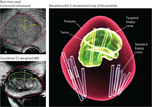
Princeton Radiology has acquired UroNav software by Invivo. With this new software we are able to gather pre-biopsy MRI images of the prostate and then seamlessly transfer them to local Urologists. Using UroNav, these Urologists can fuse the pre-biopsy MRI images with real time ultrasound-guided biopsy images for excellent delineation of the prostate and suspicious lesions, as well as clear visualization of the biopsy needle – allowing Urologists to directly aim biopsy needed at lesions. Better images means better results.

Prostate cancer is the only type of solid organ tumor that is usually diagnosed sight unseen with hit-or-miss tissue biopsies.

For decades, Urologists have used a systematic, but blind approach to sample prostate tissue in men with an elevated PSA level and no palpable lesion, taking up to 18 core needle biopsies in scattered sections of the organ. Using this method doctors can’t be sure that they haven’t missed an aggressive tumor hidden in the 99% of tissue that was not biopsied, and they sometimes end up sticking needles into men who have no tumors, only an elevated PSA level.

Now, due to the precise targeting provided by MRI, many fewer biopsy samples are needed for accurate diagnosis – sometimes 80 to 90% fewer.  Fewer needle-sticks means lower risk of infection, bleeding, pain, and means and shorter recovery time. And thanks to the recent advance called multi-parametric MRI, specially trained radiologists can gauge the aggressiveness of a prostate lesion not only by how it looks, but also by how tightly its cells are packed, how blood flows through it, and its chemical makeup.

Targeted MRI/Ultrasound biopsy is poised to become the new standard in prostate cancer detection. Princeton Radiology is proud to collaborate with local Urologists to bring the next generation of prostate care to our patients.

Prostrate Care

Image: Magnetic resonance (MR)/ultrasound fusion combines MR images of the prostate (bottom left, red line) with real-time ultrasound images of the prostate (top left, red line) to assist in targeted biopsy of a previously identified lesion (green line). The location of the biopsy can be recorded (yellow line), and a reconstructed 3-dimensional map of the prostate can be generated at the conclusion of the biopsy (right). Standard biopsy cores and targeted biopsy cores are highlighted here for comparison.

Thumbs Up to Savings!

New Forms of Radiation Can Benefit Cancer Patients

By Dr. Edward M. Soffen
Radiation oncologist, J. Seward Johnson Sr. Radiation Oncology Center, University Medical Center at Princeton
Friday, March 29, 1996

Publicity about radon, nuclear waste and fallout have caused the public to view radiation as a danger. The use of radiation in cancer therapy, however, is one instance in which its power has been harnessed to the benefit of humankind.
For many years, radioactive isotopes have been important tools in diagnosing and treating patients with cancer. Today, the therapeutic role of radiation has expanded to include two new forms of radiopharmaceutical compounds.

The first new area includes systemic radiotherapy and uses the radionuclide strontium 89, which is given intravenously to treat cancer that has spread to the bone in a process called metastasis. The second area involves new radioactive seed pellets that can be implanted in the prostate gland to control or eradicate localized prostate cancer.

Strontium 89 (under the brand name Metastron) is used to treat painful bone metastases that can accompany breast or prostate cancer. Once injected, strontium 89 is selectively taken up by the bone tissue surrounding the metastasis. The body treats the strontium as if it were calcium, putting it into the bone surrounding the cancer. The strontium then bathes the tumor with radiation while sparing all the normal tissues in the body. Strontium emits beta particles which kill the cancer. The tumor shrinks, relieving pain. The development of painful new tumor sites is also slowed or eliminated.

Previously, the palliative treatment of choice included powerful pain medications such as morphine that are derived from the opium plant. These medications mask the pain, but do not actually kill the cancer. They also have side effects that can interfere with the patient’s ability to enjoy life.

In its use at University Medical Center at Princeton and other cancer treatment centers, strontium 89 therapy has improved the quality of life for patients whose care focuses on the palliation of pain. When patients receive strontium 89 therapy, the great majority – over 75 percent – experience significant pain relief and fewer report new pain areas.

Many are able to dramatically reduce or completely eliminate their dependence on analgesics. With increased freedom from opiates, their quality of life improves dramatically. Among the lifestyle changes for these patients has been an increase in mobility that improves their ability to participate in familiar activities and return to a more normal lifestyle. After one injection, the therapy may be complete or may be repeated at three-month intervals if necessary.

Strontium 89 systemic radiotherapy is still a new tool in the fight against cancer. To date, it has been used on a selective group of patients as a valuable adjunct to the use of External Beam Radiation. EBR treats the metastatic cancer that falls within the range of the focused X-ray beam, but the strontium radioisotopes target all skeletal metastases, including those that may not yet have been diagnosed.
Like many other forms of modern cancer treatment, strontium 89 is done on an outpatient basis, further helping the patient to enjoy as normal a lifestyle as possible. Other treatments are available to control metastatic prostate cancer, including hormonal manipulation or chemotherapy. Both of these may be used before, after or in conjunction with, strontium 89.

On the other side of the treatment spectrum, the best cure for prostate cancer remains early detection. Through regular check-ups, starting at age 40, a man who develops prostate cancer may have it addressed at an earlier-therefore more curable-stage. Earlier detection and treatment may also improve the patient’s ability to retain his sexual potency.

Since prostate cancer remains one of the most common cancers among men, new treatments continue to be researched and applied. There are often excellent options available to treat early stage prostate cancer. They include radical prostatectomy, three-dimensional conformal external beam radiation and prostate seed implantation of the prostate gland.

Seed implantation within the prostate, formerly performed through open surgery, is now performed far more precisely, using ultrasound guidance. Furthermore, three-dimensional treatment planning is made possible using sophisticated new computer
software. Radioisotopes with improved properties are bringing radioactive seed implantation of the prostate gland into the mainstream of local, definitive therapy.
Unlike some other treatments for prostate cancer, the risk of impotency or incontinence with seed implantation is quite small. The ultimate decision regarding the best treatment modality for each individual patient needs to be made jointly between the patient, family members, the urologist and the radiation oncologist.

Copyright © 1996-99 The Princeton Packet, Inc.

Coronary Artery Test Provides Advance Warning

By: Dr. Jonathan Lebowitz,
University Medical Center at Princeton
July 20, 2001

According to the American Heart Association, each year 1.5 million Americans have heart attacks, and the number is rising. For 20 to 40 percent of these people, a heart attack is the first symptom of heart disease, and one in three people does not survive a first heart attack.

To help reverse this trend, it is important to take sensible steps to ensure a healthy heart before a tragedy occurs. In addition to making smart choices regarding nutrition, exercise and lifestyle, there is an exciting testing tool that can help you and your doctor assess your heart health — the coronary artery CT scan.
Coronary arteries are blood vessels that supply oxygen to your heart. Deposits called “plaque” can build up in your coronary arteries, which may eventually cause blockage. Over time, calcium can accumulate in part or all of the plaque. The more plaque that is present, the more calcium can accumulate. High levels of calcium may indicate that at least one major heart artery has a blockage, which can cause heart attacks.

The coronary CT scan evaluates the presence of — and quantifies — calcified plaque in the coronary arteries, generating a coronary artery calcification (CAC) score. This score reflects a broad estimate of cardiac risk and can be compared to average scores of people in the same age and gender category.

Regardless of the total score, it is imperative that your doctor evaluate all your risk factors. After discussion, your doctor may recommend lifestyle changes, medications, or other tests that would be appropriate.

Please note: You should not schedule this test if you are experiencing acute chest pain or other acute cardiac symptoms. Instead, call your doctor immediately.
The calcium cardiac scoring scan has been available for about 10 years, but it is only quite recently that the equipment has become faster and more accurate. The scan is done using fast spiral CT technology, without contrast, and takes less than five minutes to perform.

Another benefit is that patients may remain fully clothed, making this a comfortable and non-invasive procedure. The radiation exposure is less than that of a routine CT scan and has not been associated with harmful biological effects.

To better understand your overall heart health, familiarize yourself with the risk factors for heart disease and identify any you may have. Some risk factors,
including age, gender, family history and personal history cannot be changed.

• Age/Gender: In the middle decades of life, the risk of heart disease is three to four times higher for men than for women. For both men over 45 and women over 55, the risk gradually increases with age.

• Family History: A family history of early heart disease increases your risk.

• Personal History: The risk of a heart attack is five to seven times higher for people who have had a heart attack or stroke than for people who have not.
Controllable risk factors include:

• Smoking: Smokers are twice as likely to have a heart attack than nonsmokers.

• High Blood Pressure: High blood pressure (140/90 or higher) weakens the heart, contributing to heart disease. Reducing your blood pressure in turn reduces your risk of heart disease, stroke, kidney failure and blindness.

• High Cholesterol: Too much cholesterol in the blood can lead to clogged arteries and eventually to coronary heart disease. Opt for foods low in saturated fat and cholesterol.

• Weight: Being overweight increases your risk of high blood pressure, diabetes and heart disease. You can significantly reduce your risk by losing even 10 or 15 lbs.

• Diabetes: A person with diabetes has a two to three times higher risk of heart disease than a person who does not have diabetes. Maintain proper weight, keep active and eat a healthy diet, high in fiber and low in refined foods.

• Sedentary Life Style: Regular physical activity can cut your risk of heart disease in half and help prevent obesity, high blood pressure, high cholesterol levels and high blood sugar. It will also improve your energy level and your ability to cope with stress.

Although you may have some or all of these risk factors, modifying them will reduce your overall risk of developing heart disease. If your risk factors warrant a closer look, you can either call your hospital directly or ask your doctor to set up a cardiac calcium scan appointment for you.

The test, currently available from University Medical Center at Princeton, costs $450 and is not yet covered by insurance. Test results are mailed to both you and your doctor, at which point you can discuss appropriate treatment options, if necessary.
To be heart smart, assess your risk factors, control those you can, keep an open dialogue with your doctor, and put medical advances to work for you.

Copyright © 1996-99 The Princeton Packet, Inc.

Quiet MRI

The world is a noisy place. With Quiet MRI your exam doesn’t have to be.
Quiet MRI has up to 97% less noise and is 100% more patient-friendly.

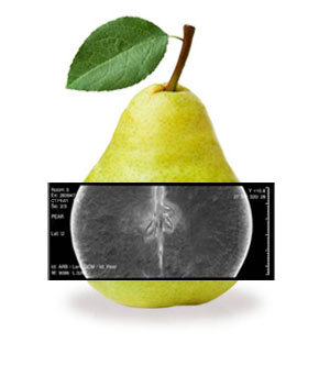
We’ve “Peared” with Siemens to Bring a Healthier CT Scan to Our Area

Our Princeton office has been “picked” by Siemens Healthcare to be a model site for their new low-dose 128-slice CT scanner.

We’ve also enhanced the CT services in our Monroe and Mercerville loctions by installing state-of-the-art low-dose 64-slice CT technology. All of our new CT scanners provide a comfortable, relaxed patient experience, and the highest image quality, while reducing harmful ionizing radiation. Patient safety is a top priority for Princeton Radiology, and our entire organization is engaged in the effort to minimize the amount of radiation used. It’s all part of our commitment to provide our patients with a healthier CT scan.

CT Pear